
Digital Cell Morphology: What It Is and Why Hematology Labs Are Switching
Ask anyone running a hematology lab about their biggest constraints, and shortage of staffing will be a frequent answer. Too many smears, too few trained eyes, and a microscope that only lets one person look at a time. Digital cell morphology helps to loosen that bottleneck – and it's now reshaping everything from routine differentials to bone marrow review.
What is digital cell morphology?
Digital cell morphology uses automated microscopy, digital imaging, and image-analysis software to help examine and classify cells. Digital cell morphology can be used for different body fluid types like blood, bone marrow and other body fluids. Rather than a technologist scanning a slide field by field under a microscope, a digital morphology analyzer automatically captures high-resolution images of cells and using artificial intelligence (AI) pre-classifies them before presenting the results to a trained professional for review.
You'll also see it called digital morphology, digital hematology or digital imaging in a broader sense. The terminology shifts depending on who you ask, but the mechanics don't: the system does the scanning, a trained professional makes the call. Morphologists still make every judgment that matters, they're just starting from a pre-classfied set of images instead of a raw slide.
How does it work?
A conventional differential means a technologist sits at a microscope, locates cells by hand, and classifies each one based on training and experience. It works, but it's slow, and quality can vary depending on who's at the scope that day.
A digital morphology system compresses that process into a few automated steps:
- The stained blood smear goes into the analyzer.
- The system scans the slide and finds the areas worth analyzing – on a blood smear this is the monolayer.
- It captures digital images of the cells.
- AI algorithms pre-classify each cell by its morphological features.
- One or several laboratory professionals reviews the pre-classified images on screen.
- They confirm or adjust the classification as needed and sign off on the final result.
Notice where the human sits in that chain – not at the start, doing the searching, but at the end, doing the judging. That's the shift.
Digital cell morphology vs. conventional microscopy
The cells being evaluated are the same. What changes is how they're found, presented, and
reviewed.
| Manual microscopy | Digital cell morphology |
| Manual slide navigation | Automated slide scanning |
| Cells located and selected manually |
Cells automatically located and digitally captured |
| Cells classified manually |
Algorithm-assisted pre-classification followed by professional review |
| Review performed at the microscope |
Review performed on a computer screen |
| Physical slide needed for conventional review |
Digital images can support remote review and consultation |
| Findings documented separately from the visual field |
Cell images and classifications can be retained with the case |
Why labs are making the switch
Labs adopt digital cell morphology because conventional microscopy has real, specific pain points – and this technology targets them directly.
It gets techs off the hunt for cells
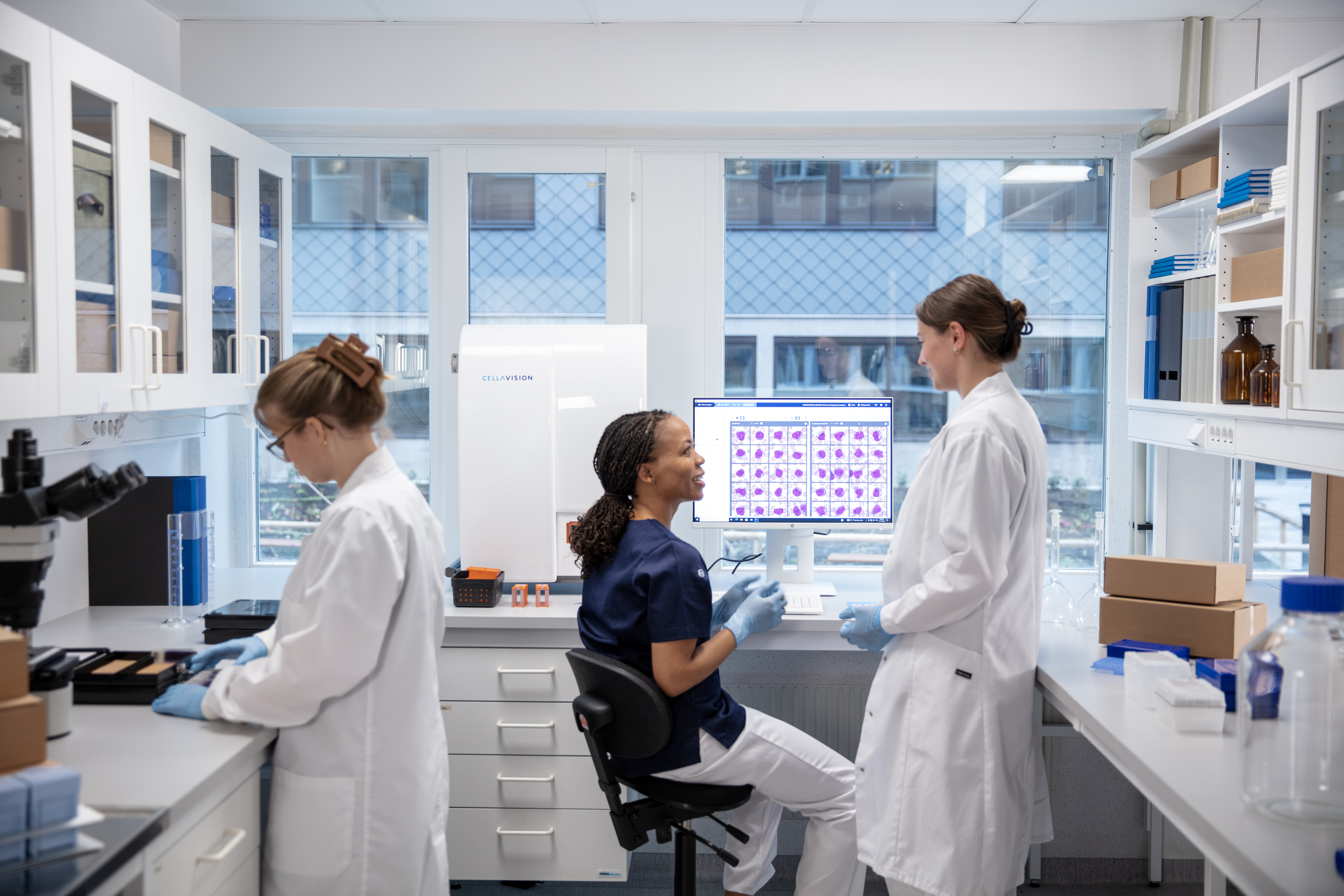
A big share of manual differential time isn't spent classifying – it's spent finding. Automating the scan and capture means technologists open a screen of pre-classified images collected in standardized and optimal locations on the smear, instead of hunting across a slide. That time gets redirected to the part of the work that requires a trained eye most.
It creates a more standardized review workflow
Manual morphology naturally involves a degree of observer-to-observer variation. A shared digital workflow gives technologists the same viewing environment, classification framework, and access to cell images across shifts, benches, and locations. [1]
Expertise stops being tied to one microscope
The image doesn't have to stay where the smear is. A tricky case in a rural lab can be pulled up by a specialist hundreds of miles away in minutes, instead of the slide going in an envelope. For networks where morphology expertise is unevenly distributed, that changes what's possible day to day. [1, 2]
It makes teaching easier
It's easier to teach from a digital image than a shared microscope. You can circle the exact cell, compare it to other cells on the same smear or on other smears, and leave it on screen as long as the conversation runs – no waiting for someone trying to relocate the same cell. [1, 3]
It makes cases easier to document and revisit
Digital workflows can retain cell images and classifications associated with a case and make the workflow steps traceable. That creates useful material for QA, research and education, consultation, and comparison with previous results. [1]
How AI is used in digital cell morphology
AI-powered image-analysis algorithms evaluate morphological features in captured digital images and use those features to suggest an initial classification of cells or other objects. They can do this rapidly and consistently across large numbers of cells. The morphologist takes it from there and confirms the call, the same way any lab result gets signed off. Speed from the algorithm, judgment from the person – that's the combination doing the work.
Where digital morphology is being used
Peripheral blood – the obvious starting point
Peripheral blood is one of the most established applications of digital cell morphology. The system locates and captures images of cells and pre-classifies them for review; many platforms are capable of classifying both white and red blood cells and provide platelet counts as well.
Body fluids – extending digital morphology beyond blood
Cell differentials in body fluids – CSF, synovial fluid, and similar samples – can run through the same kind of digital workflow, giving technologists images to classify and verify rather than requiring manual microscopy.
Digital bone marrow analysis – the next frontier
Bone marrow aspirate review is one of the most challenging applications to digitize, and for good reason: the diversity of sample preparations, cell types and maturation stages demands substantial morphology expertise.
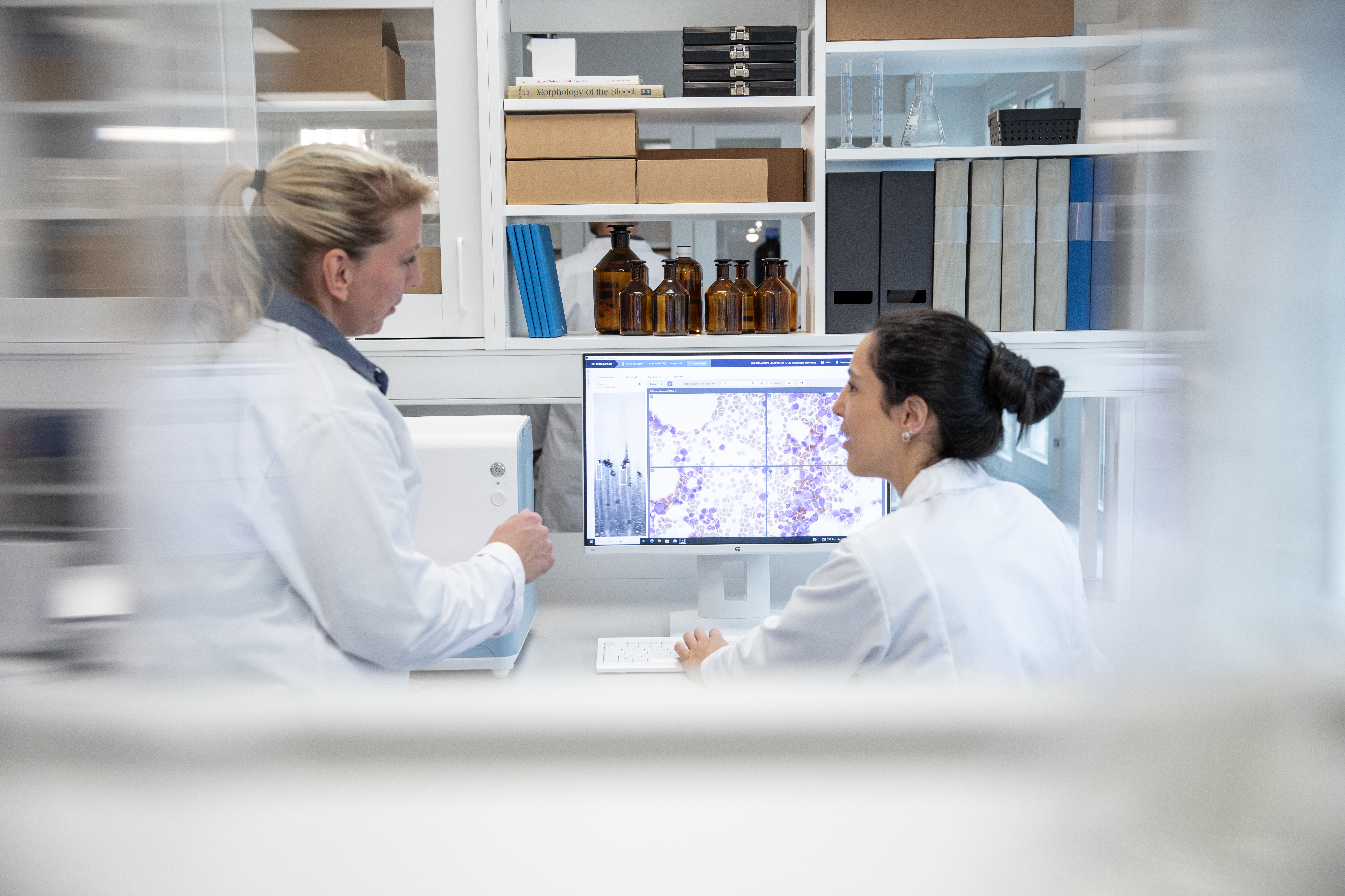
Digital bone marrow analysis brings together automated location, digital imaging, cell pre- classification, and expert review in a workflow designed for bone marrow morphology. Learn more about digital bone marrow analysis workflows →
How to choose a digital cell morphology system
The camera and analyzer are only part of the picture – the workflow and infrastructure around them matters just as much. Before committing, it's worth thinking through:
How much clinical validation stands behind the classification algorithm – published studies, not just marketing claims
- How many cell types and morphological characteristics the system classifies, across WBCs, RBCs, and platelets
- How easy it is for a technologist to reclassify a cell if needed
- Whether remote review is genuinely built for daily use, not just technically possible
- Whether it integrates with your existing LIS without a custom project
- What training is required for staff to become comfortable with the new workflow
- Whether the system supports the sample types and applications your laboratory needs today – and those you may want to add later
- What other optimized infrastructure or other support (for example for sample preparation) is connected to the analyzer itself which can speed up and increase the quality of your work
- How long the vendor has been in this space, and how stable the platform roadmap looks over the next 5–10 years
Sample volume, staffing, and existing equipment matter for finding the right fit today. Track record and validation depth can be just as important when considering how well that choice will hold up over the next five or ten years.
Frequently asked questions
What is digital cell morphology?
Digital cell morphology is the use of automated imaging and software to locate, capture, and pre-classify blood cells on a slide, so a trained technologist reviews and confirms results on screen instead of scanning every field manually.
Does this replace the technologist's job?
No – and this is a common misconception. The system pre-classifies; the human still verifies. The laboratory professional remains responsible for reviewing and verifying the classification. What changes is how much manual searching they have to do to get there.
What's the real difference from a regular microscope?
With a conventional microscope, the laboratory professional works directly at the microscope to find and classify cells. With digital cell morphology, the system captures images and pre-classifies cells, which are then presented on screen for review and verification. This changes not only how the work is performed, but also where it can be performed. Cell images can be reviewed at a workstation, revisited later, and depending on the setup, shared with other colleagues or reviewed remotely.
Is AI used to make the final decision?
No. In digital cell morphology, image-analysis algorithms can pre-classify cells and present the results to the laboratory professional for review. The professional can review the results before verification. The exact workflow and performance depend on the system and application.
The future of digital cell morphology
Less manual microscopy. More support from increasingly capable image-analysis algorithms. Morphology expertise that can be accessed beyond a single microscope.
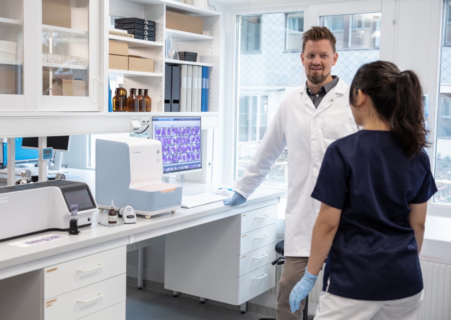
As digital morphology technology develops, the direction is toward workflows where automation handles more of the repetitive tasks like searching, imaging, and pre- classification, while laboratory professionals focus their expertise on review and interpretation. Digital collaboration also makes it easier to bring additional expertise into challenging cases.
CellaVision's approach
CellaVision builds digital microscopy systems aimed at automating the repetitive parts of morphology review without taking the decision-making out of human hands. Its solutions combine automated microscopy, image capture, and analysis software across peripheral blood, body fluids, and bone marrow aspirate applications. Explore CellaVision's digital cell morphology solutions →
References
[1] A Survey Study of Benefits and Limitations of using CellaVision™ DM96 for Peripheral Blood Differentials
[2] Improving turn-around times in low-throughput distributed hematology laboratory settings with the CellaVision® DC-1 instrument
[3] Development of a peripheral blood morphology proficiency assessment program using the CellaVision® Proficiency Software
News
CellaVision’s Digital Morphology Solutions Trusted by Top U.S. Hospitals in 2025 Rankings
CellaVision, a global leader in digital cell morphology, is proud to announce that all 20 of the top...

Celebrating 30 Years of Innovation: A Partnership with Region Skåne
As CellaVision marks its 30th anniversary, we take a moment to reflect on our journey and the...
