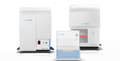
CellaVision - the essential part of every laboratory
We redefine and transform the process of performing blood differentials for labs of all sizes.
CellaVision delivers unique solutions for digital cell morphology
CellaVision offers scalable solutions adapted to the customer’s needs regarding analysis capacity, analysis type, sample preparation solutions, centralization of data and monitoring of workflow. Using analyzers, software and reagents from CellaVision laboratories can standardize and improve the efficiency and quality of your work.
Sample preparation
Sample preparation is an important part of qualitative analysis. For high-volume routine analysis, CellaVision offers a portfolio of reagents which ensures that blood smears are optimally prepared for reading by DCM. For low-volume routine analysis, sample preparation exerts a greater challenge, as the steps often rely on manual methods. CellaVision offers a semi-automatic package solution, consisting of the RAL Smearbox, RAL Stainbox and the CellaVision DC-1 together with reagents and a staining protocol specially adapted to the CellaVision DC-1. When used together, it ensures a qualitative result every time.
In addition to the conventional formulated staining solutions, the company offers a unique methanol-free product, RAL MCDh (Micro Chromatic Detection for hematology), with the additional benefit of being safer to handle in the laboratory.
CellaVision also offers RAL staining reagents in the areas of microbiology, pathology and cytology, that serve a stable customer base, and which can represent future market development
Routine analysis of blood
High-volume routine analysis is supported by CellaVision DM1200 and CellaVision DM9600 which are designed to automate, simplify and streamline the hematology workflow in large laboratories. Low-volume routine analysis is supported by CellaVision DC-1 which is designed to standardize and simplify differentiation of blood and body fluids with digital cell morphology in small and medium-sized laboratories. The systems use high speed robotics and digital image analysis technology to automatically localize and take high-quality images of cells.
Explore more
We are CellaVision

Products

Investors
