
Monthly cell challenge October 2026: Beyond the Numbers
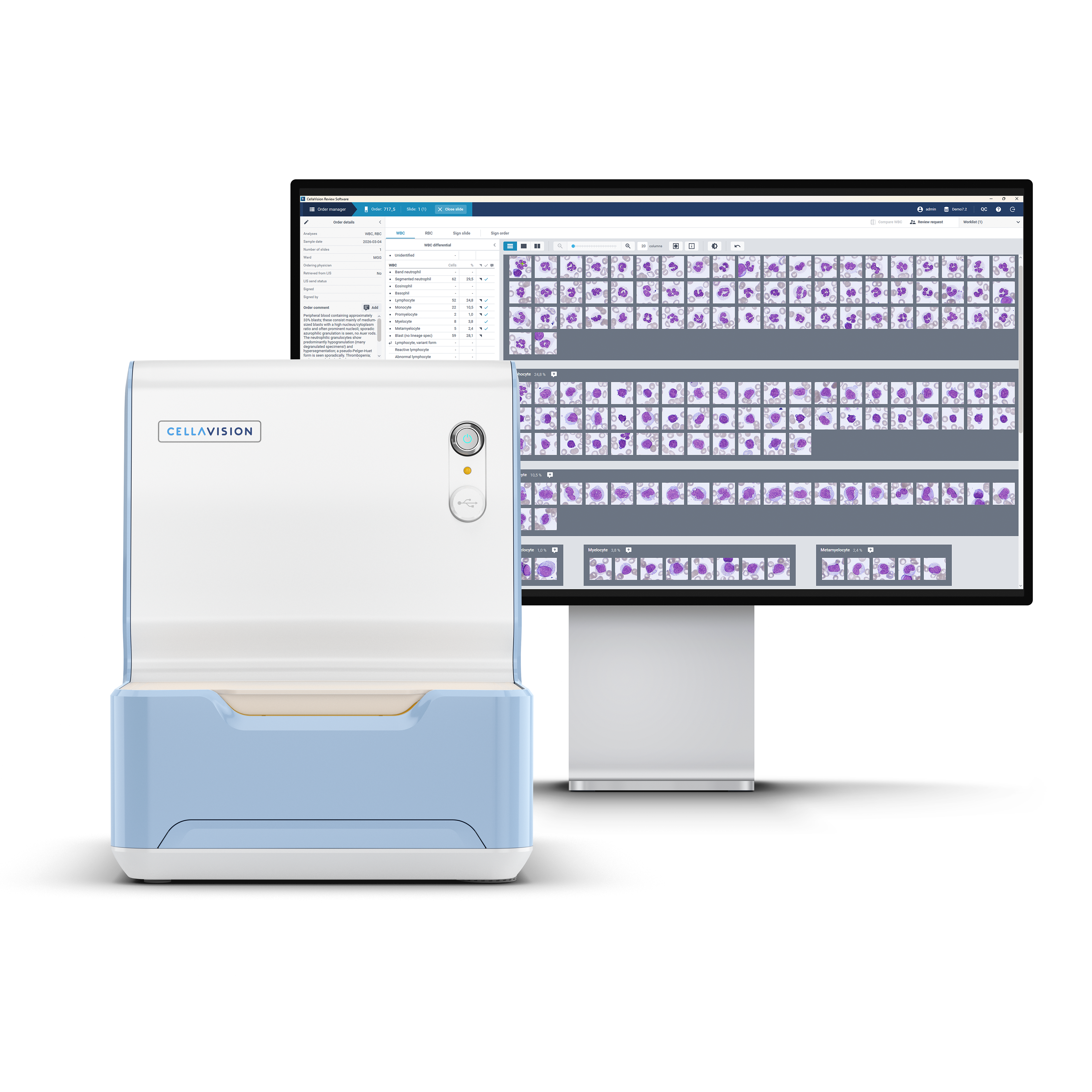
A 68-year-old man presented to his primary care physician with increasing fatigue over the past two months. He reported shortness of breath during everyday activities and noticed that he bruised more easily than usual, sometimes without any apparent injury. He also experienced occasional dizziness and a gradual decline in his ability to carry out daily activities.
On physical examination, the patient appeared pale, with several bruises visible on his arms and legs. No significant lymphadenopathy or splenomegaly was detected.
Based on his symptoms and clinical findings, a complete blood count (CBC) and peripheral blood smear examination were requested.
CBC results:
| WBC | 8,6 | 10⁹/L |
| RBC | 2,74 | 1012/L |
| HGB | 86 | g/L |
| MCV | 96 | fL |
| MCH | 31,4 | pg |
| PLT | 54 | 10⁹/L |
The CBC demonstrated moderate anemia and marked thrombocytopenia, with a white blood cell count within the normal reference range.
Blood smear analysis classification on CellaVision® DC-1
| WBC Differential | % | x109/L |
| Neutrophils | 29,5 | 2,5 |
| Lymphocytes | 24,8 | 2,1 |
| Monocytes | 10,5 | 0,9 |
| Promyelocytes | 1,0 | 0,1 |
| Myelocytes | 3,8 | 0,3 |
| Metamyelocytes | 2,4 | 0,2 |
| Blasts | 28,1 | 2,4 |
| NRBC | 3/100 WBC |
Blood smear findings
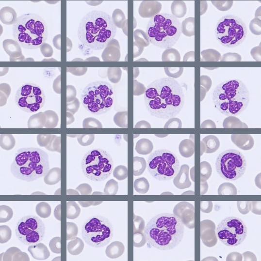
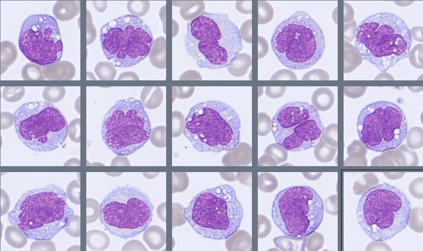
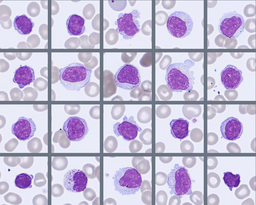
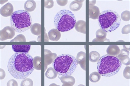
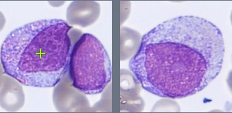
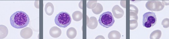
Microscopic examination of the peripheral blood smear revealed a markedly abnormal white blood cell population. The differential count showed a substantial proportion of circulating blasts (28%), together with immature myeloid cells, including promyelocytes, myelocytes, and metamyelocytes.
The blasts were predominantly medium-sized, with a high nuclear-to-cytoplasmic ratio, fine chromatin, and prominent nucleoli. Some displayed fine azurophilic granules, while no Auer rods were identified.
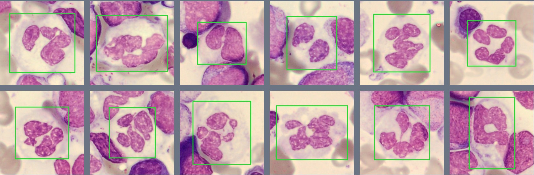
Mature neutrophils showed pronounced morphological abnormalities, including hypogranulation and abnormal nuclear segmentation. Hypersegmented neutrophils were frequently observed. Occasional nucleated red blood cells were present. Platelets were reduced in number.
The combination of numerous circulating blasts, immature granulocytes, and dysplasia triggered further investigation, including bone marrow examination and flow-cytometric immunophenotyping.
Bone marrow aspirate findings
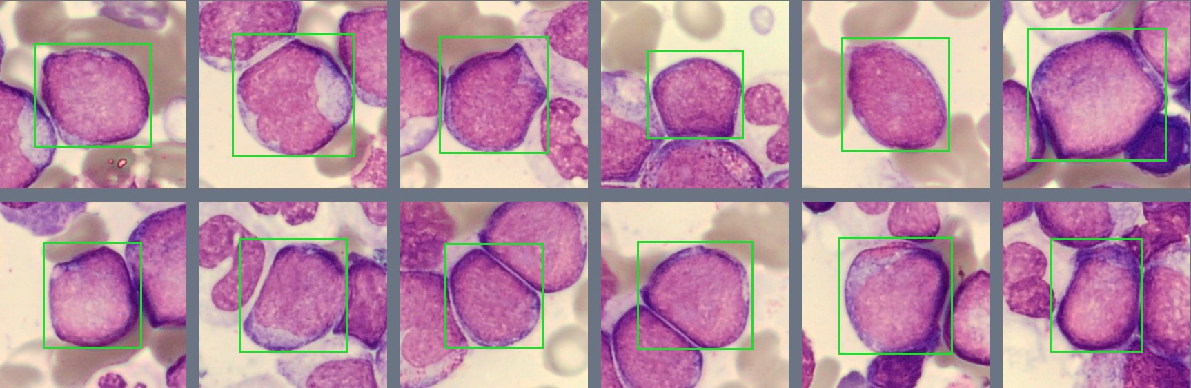
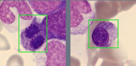
The bone marrow aspirate was of good quality and markedly hypercellular, with approximately 20% blasts. The blasts were predominantly medium-sized, with a high nuclear-to-cytoplasmic ratio and occasionally prominent nucleoli.
Dysplastic changes were observed in both the granulocytic and erythroid lineages. Granulopoiesis showed abnormal nuclear segmentation and hypogranulation, while erythropoiesis also displayed dysplastic features.
Immunophenotyping
Flow-cytometric analysis of the bone marrow identified approximately 16% CD34-positive cells. The blast population expressed CD13 and CD33, with weak MPO expression and weak, partial HLA-DR positivity. The blasts were negative for the other tested myeloid, T-cell, and B-cell markers.
Cytogenetic and molecular findings
Cytogenetic analysis of the bone marrow revealed an abnormal karyotype with monosomy 7 detected in 12 of 20 analyzed metaphases. No additional AML-defining genetic abnormalities were identified by molecular testing.
Diagnosis
Acute Myeloid Leukemia, Myelodysplasia-Related (AML-MR).
Discussion
Acute myeloid leukemia, myelodysplasia-related (AML-MR), is a subtype of acute myeloid leukemia characterized by an increased number of immature myeloid cells together with specific genetic abnormalities or a history of myelodysplastic neoplasms (MDS) or MDS/myeloproliferative neoplasms (MDS/MPN) [1][2]. AML-MR mainly affects older adults and is often associated with impaired blood cell production. Common symptoms include fatigue, shortness of breath, easy bruising, bleeding, and recurrent infections, usually caused by anemia, thrombocytopenia, or reduced numbers of functional white blood cells [2].
The peripheral blood smear may show circulating blasts, often accompanied by dysplastic changes in other blood cells. Myeloid blasts are typically medium to large cells with a high nuclear-to-cytoplasmic ratio, fine chromatin, and visible nucleoli. Some may contain azurophilic granules or Auer rods, although these are not always present. Neutrophils may show reduced granulation or abnormal nuclear segmentation, while red blood cells can display variation in size and shape. These morphological findings may suggest disturbed blood cell development but are not specific to AML-MR [1][3].
Bone marrow examination typically reveals an increased proportion of blasts, often with dysplastic changes affecting one or more hematopoietic lineages. The marrow is frequently hypercellular, although cellularity may vary. Flow cytometry is important for identifying and characterizing the blast population. Myeloid blasts commonly express markers such as CD13, CD33, CD34, CD117, and MPO, although the expression pattern varies between patients. Immunophenotyping supports the diagnosis of AML but cannot independently establish the AML-MR subtype [2][3].
According to the WHO Classification of Haematolymphoid Tumours (2024), AML-MR is defined by specific myelodysplasia-related genetic abnormalities or a documented history of MDS or MDS/MPN. A blast count of at least 20% in the peripheral blood or bone marrow is generally required. Importantly, multilineage dysplasia alone is no longer sufficient for the diagnosis of AML-MR [1][4].
Cytogenetic and molecular testing therefore play a central role in classification. Myelodysplasia-related chromosomal abnormalities include monosomy 7, deletion of chromosome 7q, and complex karyotypes. Mutations in genes such as ASXL1, SRSF2, SF3B1, U2AF1, and RUNX1 may also support the diagnosis. These findings help distinguish AML-MR from other AML subtypes and provide important information about prognosis and treatment decisions [1][4].
AML-MR is generally associated with an unfavorable prognosis compared with many other AML subtypes, although outcomes depend on the patient's age, overall health, genetic findings, and response to treatment. Treatment options include intensive chemotherapy, lower-intensity regimens such as hypomethylating agents combined with venetoclax, and allogeneic stem cell transplantation in eligible patients. Targeted therapies may also be considered when suitable molecular abnormalities are present [2].
In this case, peripheral blood and bone marrow analysis using CellaVision® DC-1 revealed an increased proportion of blasts, along with immature granulocytes and pronounced dysplastic changes. Flow-cytometric immunophenotyping supported the myeloid origin of the blasts. Cytogenetic analysis identified monosomy 7, a myelodysplasia-related chromosomal abnormality, confirming the diagnosis of AML-MR according to WHO 2024.
This case demonstrates the importance of combining morphological examination with immunophenotyping and genetic testing. While blood smear findings may raise suspicion of AML, cytogenetic and molecular investigations are essential for accurate classification and can provide valuable guidance for further clinical management.
References
[1] WHO Classification of Tumours Editorial Board. Haematolymphoid Tumours. WHO Classification of Tumours, 5th ed. Vol. 11. Lyon (France): International Agency for Research on Cancer; 2024.
[2] Döhner H, Wei AH, Appelbaum FR, Craddock C, DiNardo CD, Dombret H, et al. Diagnosis and management of AML in adults: 2022 recommendations from an international expert panel on behalf of the ELN. Blood. 2022;140(12):1345–1377. doi:10.1182/blood.2022016867.
[3] Bain BJ. Blood Cells: A Practical Guide. 6th ed. Hoboken (NJ): Wiley-Blackwell; 2021.
[4] Hörst K, Kühn C, Haferlach C, Haferlach T, Khoury JD. Genetic studies in clonal haematopoiesis, myelodysplastic neoplasms and acute myeloid leukaemia – a practical guide to WHO-HAEM5. Med Genet. 2024;36(1):21–29. doi:10.1515/medgen-2024-2010.