Monthly Cell Challenge #06 / 2026
The Racing Pigeon's Hidden Passenger
A 2-year-old racing pigeon (Columba livia domestica) was presented to a veterinarian after its owner had noticed a gradual decline in flight performance over the past month. The bird showed reduced endurance during training sessions, appeared less active than the other pigeons in the loft, and had lost weight over time. On physical examination, the pigeon was found to be in slightly poor body condition and had mildly pale mucous membranes. No other significant abnormalities were detected.
A blood sample was collected for a hematologic evaluation. Due to logistical delays, the sample was not analyzed immediately and remained stored for an extended period before blood smear evaluation.
CBC results:
| Test | Result | Units | Ref |
|---|---|---|---|
| WBC | 18.5 | x109/L | 9.0 – 17.0 ×10⁹/L |
| RBC | 2.1 | 1012/L | 2.5 – 4.5 ×10¹²/L |
| HCT | 0.29 | L/L | 0.35 – 0.55 L/L |
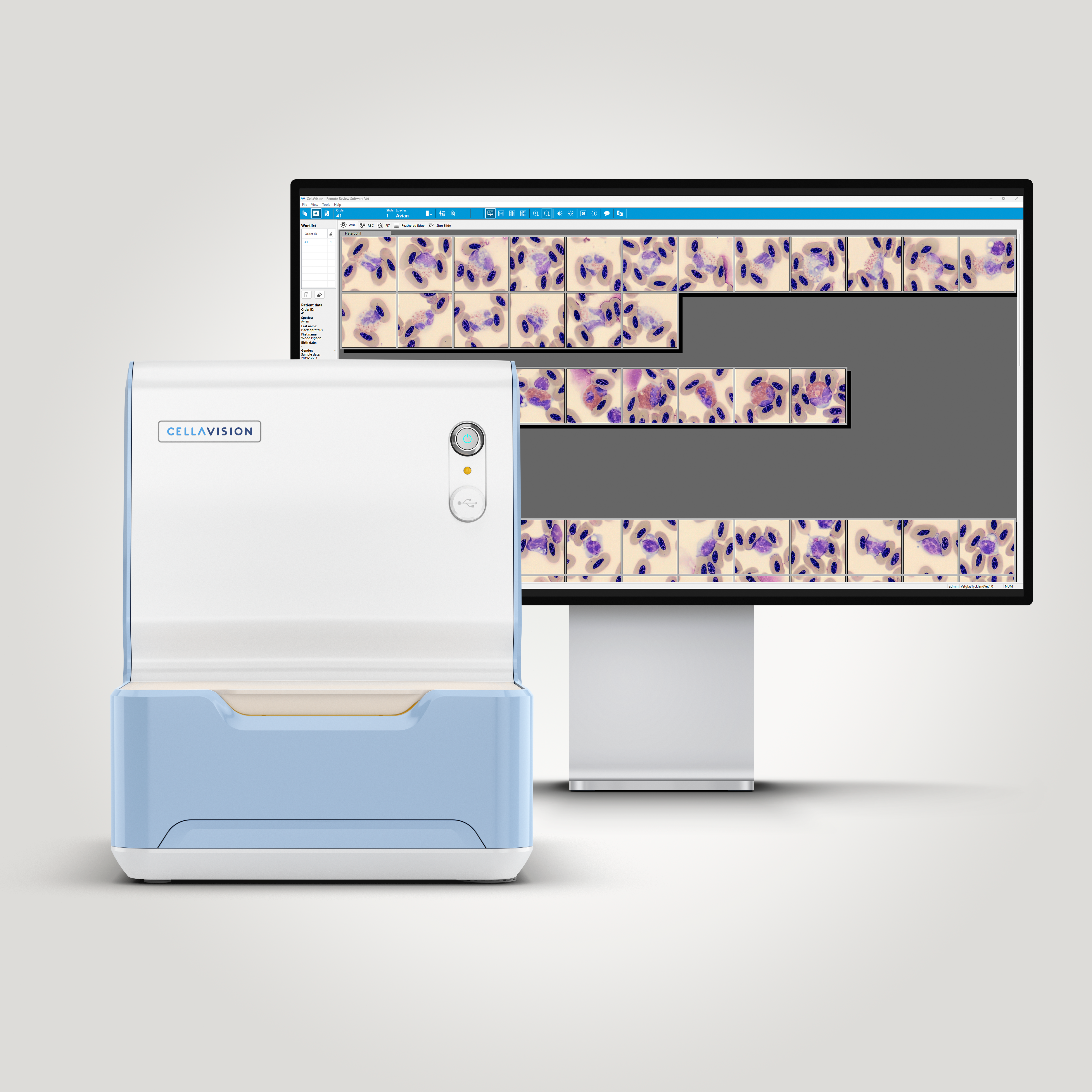
Smear review on CellaVision® DC-1 VET Remote review
| WBC Differential | % | x109/L | Ref |
|---|---|---|---|
| Heterophils | 22.5 | 4.2 | 1.5 – 7.0×10⁹/L |
| Eosinophils | 11.3 | 2.1 | 0 – 1.5×10⁹/L |
| Basophils | 2.5 | 0.5 | 0 – 0.5×10⁹/L |
| Lymphocytes | 48.8 | 9.0 | 3.0 – 10.0×10⁹/L |
| Monocytes | 15.0 | 2.8 | 0 – 1.5×10⁹/L |
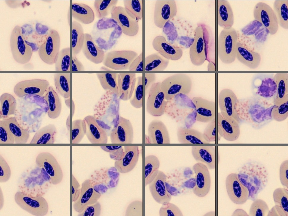
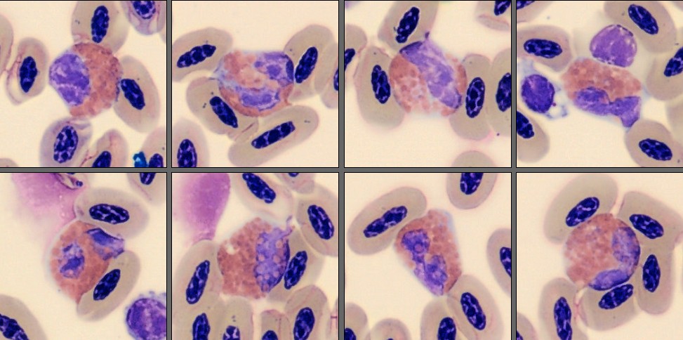
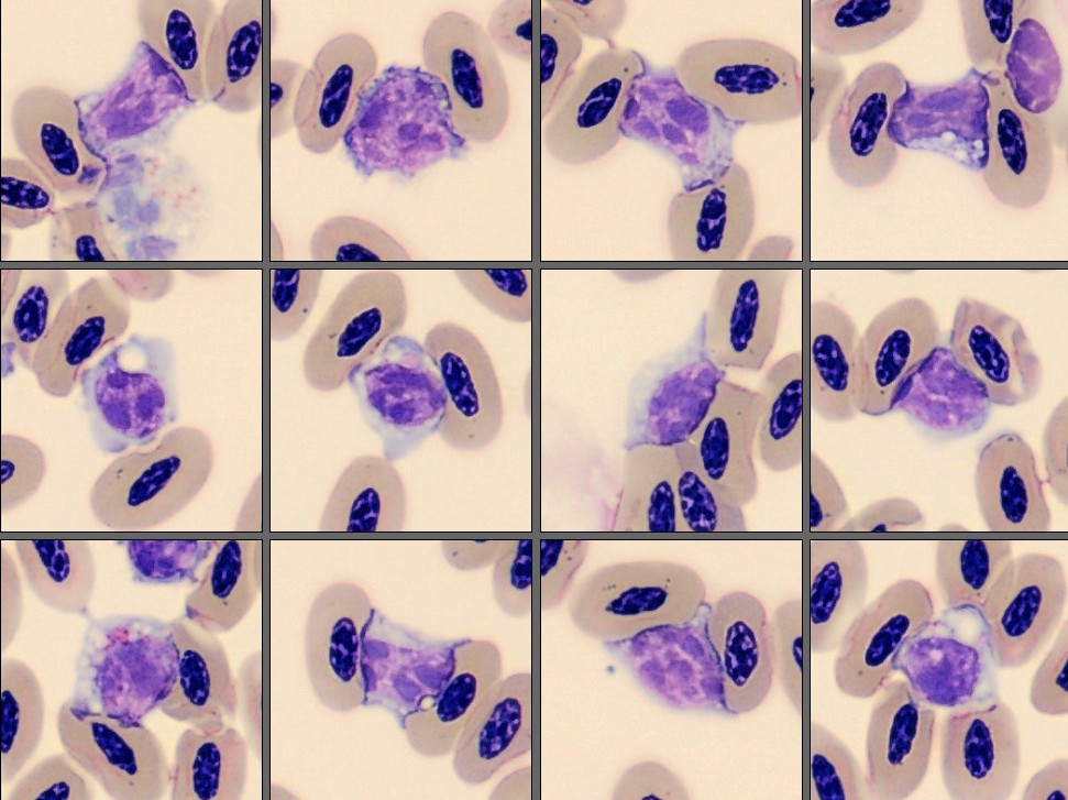
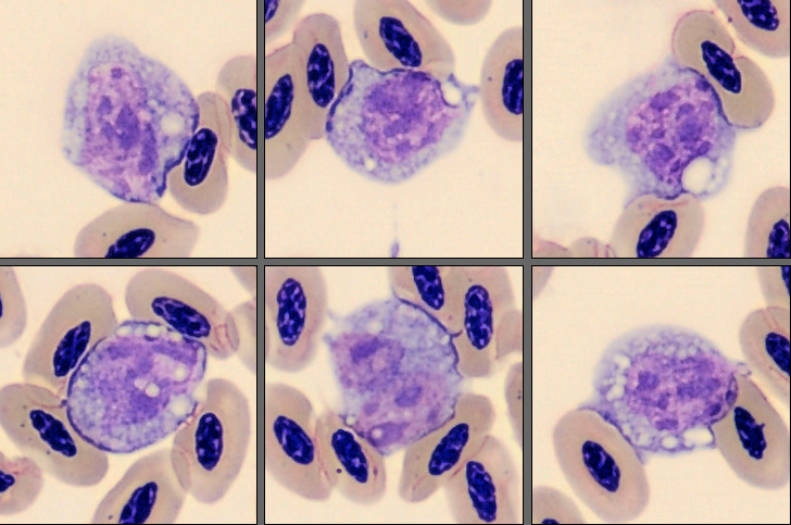
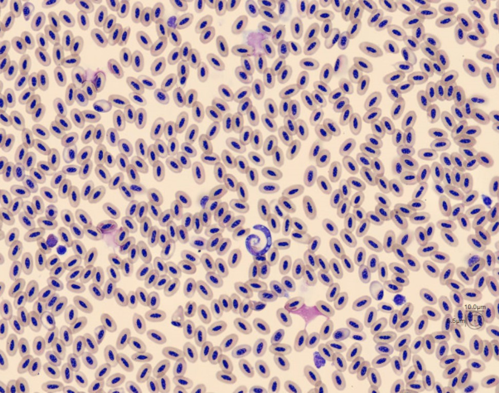
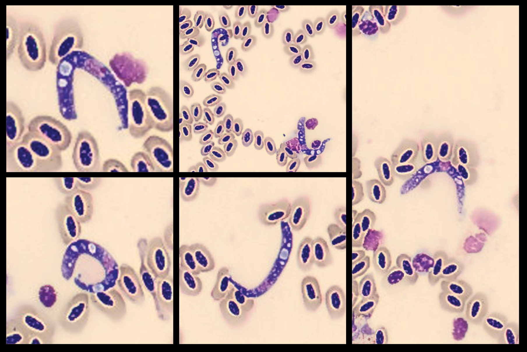
The leukocyte morphology appeared largely unremarkable, although mild eosinophilia and monocytosis were noted. The erythrogram indicated mild to moderate anemia. During the review of the WBC gallery, numerous unusual, elongated structures were observed. These were pre-classified as smudge and artefacts and appeared extracellular, varied in size, and contained prominent vacuole-like areas. Similar structures were present in the RBC overview and in even greater numbers in the feathered edge scan. The appearance of these structures was not immediately recognizable and represented the most striking finding in the smear.
This case also highlights the importance of reviewing all avalible image galleries during blood smear evaluation.
Diagnosis:
Haemoproteus columbae Infection
Discussion:
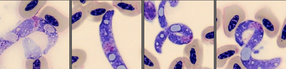
The important aspect of this case was that the blood sample was not examined immediately after collection. Following prolonged storage before microscopic evaluation, the parasites exhibited marked morphological changes and no longer resembled the classic intraerythrocytic gametocytes most laboratorians associate with Haemoproteus columbae [1][2][3]. When it comes to Haemoproteus spp., extracellular developmental stages are normally found in host tissues and organs, whereas circulating blood stages are typically observed within erythrocytes [1][2][3].
Haemoproteus columbae is one of the most common haemosporidian parasites of domestic pigeons worldwide and is transmitted by the pigeon louse fly (Pseudolynchia canariensis) [1][4]. Infection is highly prevalent in both feral and racing pigeons and is often subclinical in adult birds. Clinical disease is more likely to occur in young birds, heavily parasitized individuals, or birds exposed to stress such as racing, transport, breeding, or concurrent illness [1][4]. When present, clinical signs are typically nonspecific and may include reduced flight performance, exercise intolerance, lethargy, weight loss, decreased appetite, and poor body condition [1][4].
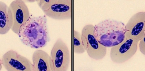
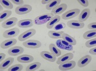
In freshly prepared blood smears, Haemoproteus parasites are usually identified as pigmented intraerythrocytic gametocytes that partially surround the erythrocyte nucleus. [1][2][3][4]. In this case, however, the organisms appeared markedly different. Instead of the typical intraerythrocytic forms, numerous elongated extracellular structures with variable shape and prominent vacuole-like areas were observed, particularly in the feathered edge of the smear.
The atypical appearance complicated the microscopic interpretation and broadened the differential diagnosis. Degenerative changes associated with sample storage can affect both blood cells and parasites, making morphological identification more challenging [2][3]. As a result, the parasite did not exhibit the typical morphology of Haemoproteus species commonly illustrated in textbooks and other reference materials.
Although the parasites lacked their characteristic morphology, infection with Haemoproteus columbae was confirmed by PCR. The atypical appearance was therefore considered most likely to be the result of sample aging rather than infection with another hemoparasite species. Morphological variation in avian hemoparasites has been linked to factors such as pre-analytical factors, parasite developmental stage, parasite burden, host-related influences, and sample quality. These factors can make morphological diagnosis challenging, particularly when blood smears are not examined shortly after collection [1][4].
Previous studies have shown that H. columbae infection may influence hematologic parameters, particularly in chronically infected birds. Reported findings include anemia, alterations in erythrocyte indices, and changes in leukocyte populations, although the severity varies considerably among individuals [5]. The mild anemia, eosinophilia, and monocytosis observed in this case was consistent with findings reported in Haemoproteus infected pigeons.
Treatment is generally considered unnecessary in clinically normal birds. Management is primarily focused on minimizing stress, optimizing husbandry, and reducing exposure to the pigeon louse fly (Pseudolynchia canariensis), the parasite's vector [1]. In clinically affected birds, antiprotozoal treatment has been described, although evidence for complete parasite clearance remains limited, and recommendations vary between studies [1][4].
This case illustrates how parasites do not always present in their textbook form. Careful smear examination, awareness of preanalytical factors, and an understanding of avian hemoparasite biology remain essential for accurate diagnosis. Even when morphology is atypical, Haemoproteus infection should remain a differential diagnosis in pigeons and other avian species [1][2][3].
References:
[1] Valkiūnas G. Avian Malaria Parasites and Other Haemosporidia. Boca Raton: CRC Press; 2005.
[2] Campbell TW, Ellis CK. Avian and Exotic Animal Hematology and Cytology. 4th ed. Ames: Wiley-Blackwell; 2013.
[3] Weiss DJ, Wardrop KJ, editors. Schalm's Veterinary Hematology. 7th ed. Ames: Wiley-Blackwell; 2022.
[4] Prompiram P, Mongkolphan C, Poltep K, et al. Baseline study of the morphological and genetic characteristics of Haemoproteus parasites in wild pigeons (Columba livia) from paddy fields in Thailand. Int J Parasitol Parasites Wildl. 2023;21:65–73.
[5] Radfar MH, Fathi S, Asl EN, Dehaghi MM, Seghinsara HR. Effect of Haemoproteus columbae infection on the hemogram of the domestic pigeon (Columba livia domestica). J Parasit Dis. 2016;40(4):1432–1435.