AI & Innovation at CellaVision: 30 Years of Intelligent Microscopy
Artificial intelligence isn’t new at CellaVision – it’s been at the core of our mission for over three decades. Long before AI became a global buzzword, our founders envisioned using it to transform the way blood cells are classified. Today, with interest in AI at an all-time high, we’re often asked how we apply it in our products and what it means for digital cell morphology.
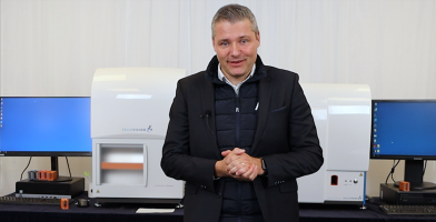
In this article, Adam Morell, responsible for devices and software at CellaVision, shares how AI has shaped our journey, how we use it today, and where the technology is headed.

How did it all begin 30 years ago?
We started working with machine learning, or AI, already in the '90s. Back then there were companies using AI for other applications, but it was rather new to use AI for this analysis - for classification of blood cells. In the beginning, we wrote our own libraries for classification based on traditional neural networks. We tailor-made specific features to distinguish between the different cell classes. With the introduction of deep learning feature design isn't important anymore since we can now feed entire images to the AI. This is just one example of how AI has evolved over the years we've been working with it. Throughout the years, we built considerable knowledge within this field, and we have successfully incorporated the latest developments into our products.
How do we use AI today?
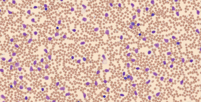
We use AI for several different tasks in our products. The most obvious ones are for the pre-classification of white blood cells and the pre-characterization of red blood cell morphologies.
Another quite unique innovation is that we use AI to acquire focused images. Traditional methods for focusing are based on acquiring many images and then choosing the best one a process which can be quite slow. Thanks to our AI based absolute focus method we can acquire focused images much faster. Essentially, we can determine whether an image is focused or not and if it's not focused, we can also determine how to reposition the analyzer to acquire a focused image. This method has significantly improved the speed of our analyzers. It is not used by others - it's unique and patented by CellaVision.
That is an excellent question. One which I'm actually asked quite often. The short answer is no. Our analyzers do not change the pre-classification once they've left CellaVision. When we develop new software, we teach the AI to classify blood cells. We then perform an extensive validation making sure that the pre-classification performs well on a large set of different samples from different labs. It wouldn't be difficult to let the AI adapt using feedback from the users, however, the AI would then start to deviate from its original state, and we could no longer make any guarantees about the results. Therefore, the AI is not learning in the lab.
What challenges do you see with AI in digital morphology?
The most challenging aspect of AI is having high-quality ground truth data. Actually, the AI itself isn't that difficult anymore, thanks to the development in recent years, there are readily available tools for anyone who wants to experiment with AI. Of course, if you want really good results you need a certain amount of knowhow. But no matter how good you are the quality of the data used to train the AI limits how good it can become.
The size of the data set used to train the AI is, of course, important. That's why we have large libraries of both slides and images at CellaVision, but even more important than size is the variability of the data. To make the AI robust, one needs to make sure that the dataset covers every variation that the AI can be subjected to. We have structured ways of making sure that all variations are covered - from staining variations to system parameters. The quality of the ground truth classification is, of course, also crucial and we make use of a wide network of top-tier experts, and each cell is classified by several experts.
How can you trust the pre-classification?
We always do a proper validation. Typically including 12 different clinical studies. The studies include about 8 million cells from 6,000 samples. The studies are also performed in different parts of the world, making sure we meet the strict regulatory requirements of highly controlled markets such as China, the EU, and the US.
What’s coming next?
We have already improved the workflow. In the hematology lab, quite a bit but I think there is more to be done. One possibility would be to remove the requirement that the user has to review each cell. Instead, the analyzer is trusted to determine the classification of some cells without any human intervention. This could dramatically reduce the workload in the lab. Another opportunity would be to integrate even more information from other instruments in the lab like, for example, the cell counters. This could provide more information to the physician and offer even greater diagnostic value. CellaVision is at the forefront of advancing laboratory technology using the latest advancements in AI.
We will continue to drive innovation, setting new standards for intelligent microscopy.
News
Why Smudge Cells Should Not Be Overlooked in the Differential of Digital Cell Morphology Systems
Smudge cells, also known as basket cells, are distorted white blood cells often caused by mechanical...

World Creativity and Innovation Day 2024
CellaVision’s long-term success is a result of our commitment to innovation. Our goal is to provide...
