
Monthly cell challenge – September 2026: Beneath the Surface
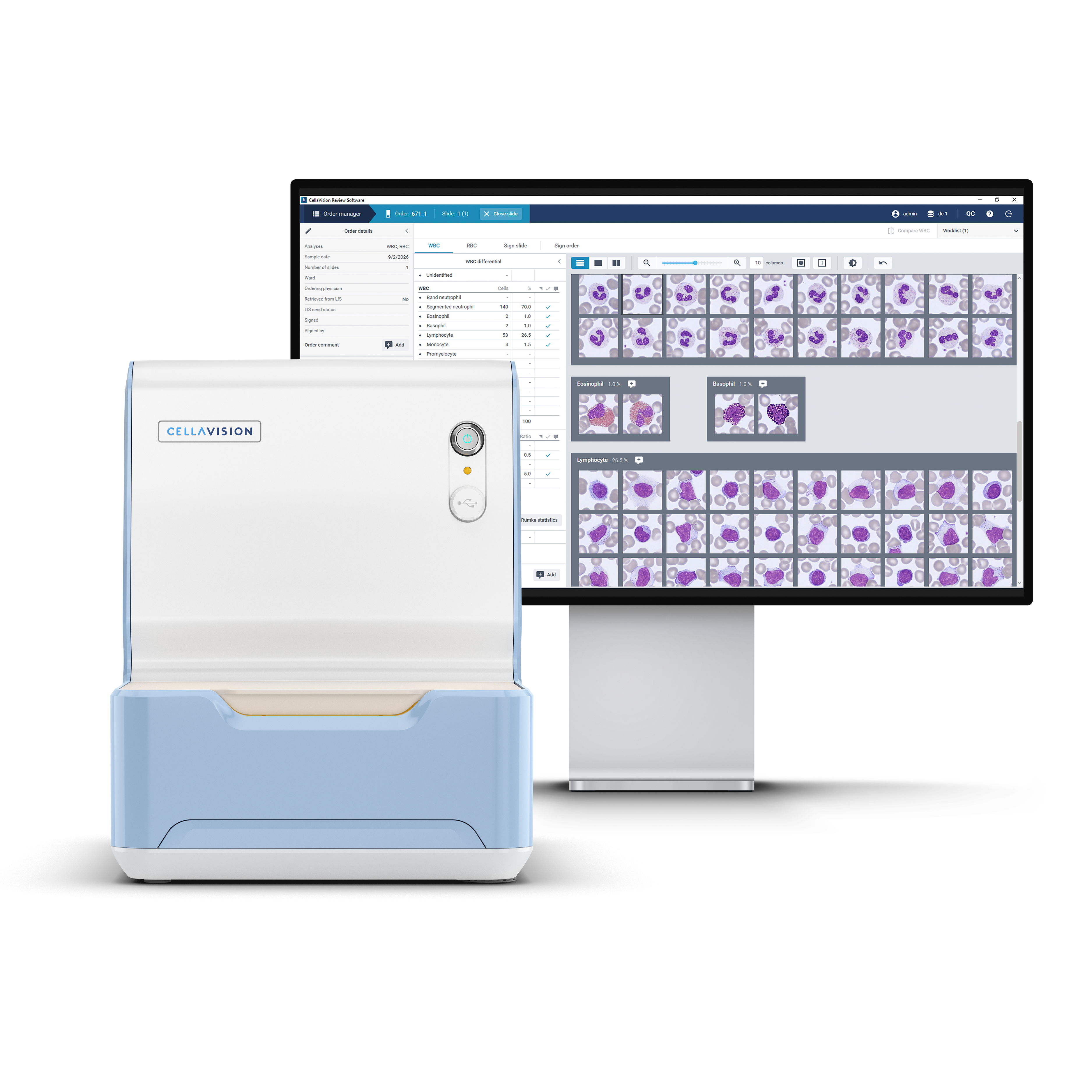
A 62-year-old man presented to his primary care physician with progressive fatigue, reduced exercise tolerance, and a sensation of fullness in the left upper abdomen. During the last months, he had experienced several respiratory infections and noticed that he bruised more easily than usual. He denied overt bleeding, fever, night sweats, and unintentional weight loss.
His medical history included well-controlled hypertension. He was not taking anticoagulant medication and had no known previous hematological disorder.
On physical examination, the patient appeared mildly pale. The spleen was palpable below the left costal margin, while no clinically significant peripheral lymphadenopathy was detected. Because of his symptoms and the clinical findings, the physician initiated further investigation.
As part of the investigation, blood samples were obtained for routine laboratory testing.
CBC results:
| Test | Result | Units |
| WBC | 2,4 | 10⁹/L |
| RBC | 3,45 | 1012/L |
| HGB | 104 | g/L |
| MCV | 93 | fL |
| MCH | 30 | pg |
| PLT | 82 | 10⁹/L |
The CBC demonstrated mild normocytic anemia, leukopenia, and thrombocytopenia.
Blood smear analysis classification on CellaVision® DC-1
| WBC Differential | % | x109/L |
| Neutrophils | 70,0 | 1,68 |
| Lymphocytes | 26,5 | 0,64 |
| Monocytes | 1,5 | 0,04 |
| Eosinophils | 1,0 | 0,02 |
| Basophils | 1,0 | 0,02 |
Blood smear findings
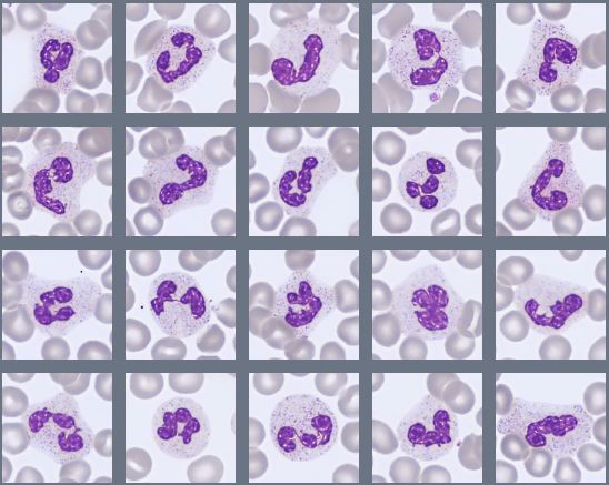
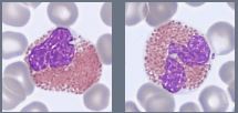
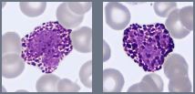
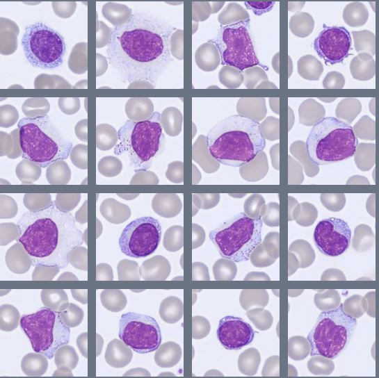
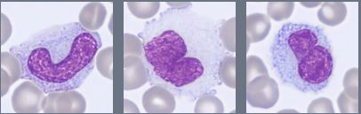
Microscopic examination of the peripheral blood smear initially showed no striking abnormality. There was no lymphocytosis, and most leukocytes displayed unremarkable morphology. Mature neutrophils showed no significant dysplastic features. Occasional eosinophils and basophils were present and displayed normal morphology. Platelets were decreased in number, with sporadic macrothrombocytes. However, a more detailed review revealed a small population of unusual lymphocyte-like cells.
These cells were predominantly small to medium-sized, with round to oval nuclei, relatively mature chromatin, and inconspicuous nucleoli. They contained a moderate amount of pale blue-gray cytoplasm. On closer examination, several cells showed fine, irregular, hair-like projections extending from the cytoplasmic border. The abnormal cells were sparse and could easily have been overlooked during the initial assessment.
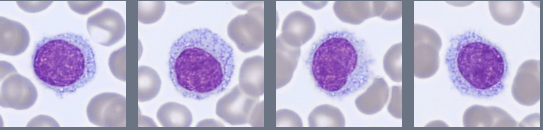
Although the abnormal lymphoid population was small, the combination of persistent cytopenias and the distinctive cytoplasmic projections raised suspicion of an underlying mature B-cell lymphoproliferative disorder. The patient was therefore referred for further evaluation, including bone marrow aspiration and flow-cytometric immunophenotyping.
Bone marrow aspirate findings
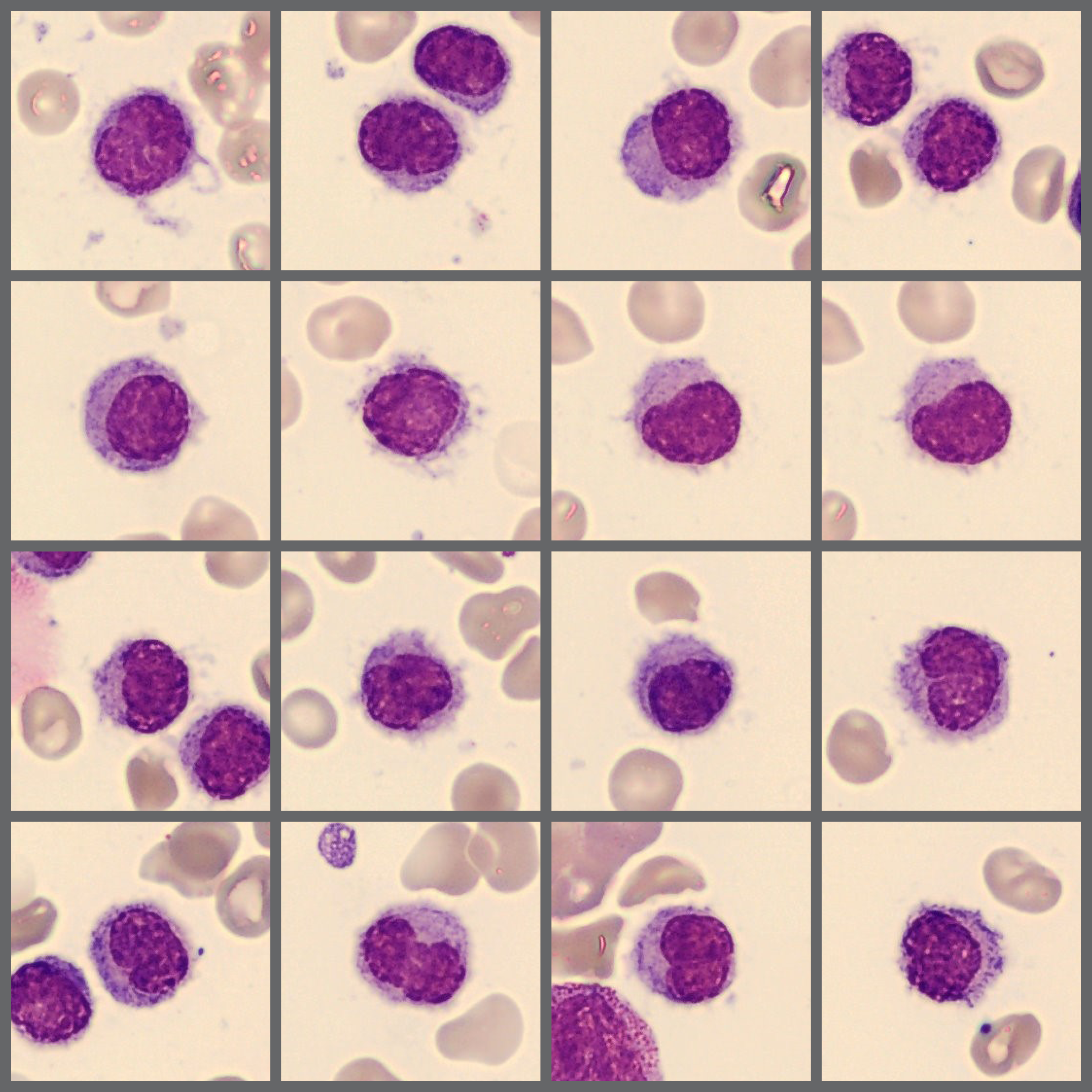
The bone marrow aspirate was of good quality and moderately cellular, with a marked increase in lymphoid cells. The abnormal lymphocytes were predominantly small to medium-sized, with round, oval, or slightly indented nuclei, moderately condensed chromatin, and inconspicuous nucleoli. They contained a moderate amount of pale blue-gray cytoplasm, often displaying fine, irregular cytoplasmic projections. The abnormal cells accounted for approximately 26% of the marrow cells and corresponded morphologically to those observed in the peripheral blood.
The remaining hematopoietic lineages showed preserved maturation without significant dysplasia or an increase in blasts.
Immunophenotyping
Flow-cytometric analysis of the bone marrow demonstrated a substantial lambda light chain-restricted monoclonal B-cell population, representing approximately 50% of the total cell population. The abnormal cells strongly expressed CD19, CD20, CD11c, and CD103, while CD5 was negative. No increase in CD34-positive blast cells was identified.
Diagnosis
Hairy Cell Leukemia (HCL).
Discussion
Hairy cell leukemia (HCL) is a rare, indolent mature B-cell neoplasm that primarily involves the bone marrow, spleen, and peripheral blood [1]. It occurs mainly in middle-aged and older adults and is considerably more common in men [1][2]. Patients frequently present with fatigue, recurrent infections, easy bruising, or abdominal discomfort caused by splenomegaly. Common laboratory findings include leukopenia, anemia, thrombocytopenia, and characteristic monocytopenia [2][4].
Hairy cells are small to medium-sized mature lymphoid cells with round, oval, or occasionally reniform nuclei, relatively mature chromatin, and inconspicuous nucleoli. They contain a moderate amount of pale blue-gray cytoplasm with fine, irregular projections that produce the characteristic “hairy” appearance [1][2]. These projections may be subtle or poorly preserved, particularly when only a few neoplastic cells are present. Careful review of the optimal monolayer and standardized morphological reporting are therefore important [3].
Morphology alone is not enough for definitive diagnosis because HCL may resemble other mature splenic B-cell neoplasms. Flow cytometry typically demonstrates a monoclonal B-cell population expressing CD19, strong CD20, CD11c, CD25, CD103, and CD123, while CD5 and CD10 are generally negative [1][4]. The BRAF V600E mutation is present in the great majority of classic HCL cases and provides valuable diagnostic support. Its absence should prompt consideration of alternative splenic B-cell neoplasms [1][4].
Bone marrow involvement is characteristic and may be interstitial, patchy, or diffuse. Increased reticulin fibrosis is common and can result in a difficult or unsuccessful aspiration. When adequate aspirate material is obtained, the abnormal cells usually correspond morphologically to those identified in peripheral blood [1][2].
In this case, the peripheral blood smear initially appeared relatively unremarkable because leukopenia was present and the abnormal cells were sparse. Upon digital review using CellaVision® Review Software a small population of mature lymphoid cells with pale cytoplasm and fine cytoplasmic projections were found. The bone marrow aspirate subsequently demonstrated marked lymphocytosis, including approximately 26% morphologically recognizable hairy cells. Flow cytometry identified a substantial lambda-restricted B-cell population with strong expression of CD19, CD20, CD11c, and CD103 and absence of CD5. The combined morphological and immunophenotypic findings supported the diagnosis of classic HCL [1][4].
Treatment is generally indicated for symptomatic cytopenias, recurrent infections, symptomatic splenomegaly, or other disease-related complications. Purine nucleoside analogues, particularly cladribine and pentostatin, are established treatments with high response rates. Rituximab and BRAF-targeted therapy may be considered in selected patients, especially in relapsed or refractory disease [4].
This case demonstrates the importance of careful blood film review in patients with otherwise unexplained cytopenias. Even a small population of abnormal lymphoid cells may provide the first indication of HCL and lead to appropriate bone marrow, immunophenotypic, and molecular investigations.
References
[1] WHO Classification of Tumours Editorial Board. Haematolymphoid tumours. 5th ed. Vol. 11. Lyon (France): International Agency for Research on Cancer; 2024.
[2] Bain BJ. Blood cells: a practical guide. 6th ed. Hoboken (NJ): Wiley-Blackwell; 2021.
[3] Palmer L, Briggs C, McFadden S, Zini G, Burthem J, Rozenberg G, et al. ICSH recommendations for the standardization of nomenclature and grading of peripheral blood cell morphological features. Int J Lab Hematol. 2015;37(3):287–303. doi:10.1111/ijlh.12327.
[4] Grever MR, Abdel-Wahab O, Andritsos LA, Banerji V, Barrientos J, Blachly JS, et al. Consensus guidelines for the diagnosis and management of patients with classic hairy cell leukemia. Blood. 2017;129(5):553–560. doi:10.1182/blood-2016-01-689422.