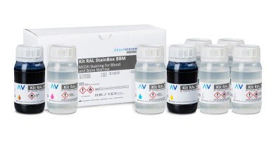
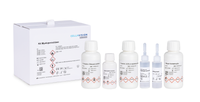
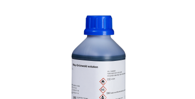
May-Grünwald Giemsa staining
Description
May-Grünwald Giemsa staining
What is MGG staining?
Mainly used in hematology, the May-Grünwald Giemsa stain is a Romanowsky type stain. It highlights medullary cells and blood cells thanks to two stains: May-Grünwald and Giemsa. The MGG staining allows to differentiate white blood cells: neutrophils, eosinophils, basophils, lymphocytes and monocytes as well as erythrocytes. This stain according to Pappenheim can also be used for tissue sections in histology.
How does MGG staining work?
May Grünwald and Giemsa are neutral stains. They are not active in alcoholic media and act selectively only when released into a buffered aqueous solution. This release induces a precipitation of the neutral stains. May Grünwald is an acidic stain while Giemsa is a basic stain. May Grünwald is composed of methylene blue and eosin. Methylene blue stains nuclei (DNA and RNA/chromatin structure), basophilic granulations, cytoplasm of lymphocytes and monocytes as well as cytoplasm proteins blue. Eosin stains red blood cells (hemoglobin), basophilic and eosinophilic granulations and neutrophilic cytoplasm proteins in red orange. Giemsa is composed of methylene azure. It stains the nuclei (DNA and chromatin structure) and the azurophilic granulations of the cytoplasm in purple.
Benefits
Produce a reliable result
Guarantee an optimal result with our RAL buffer, which allows for better rinsability and a cleaner slide base. Our May Grünwald and Giemsa stains are of consistent quality due to the synthesis of the stain powders in our own factory.
These products are CE/IVDR marked.
Information Request
Want to learn more about our product concept, request a demonstration, or just get in touch with CellaVision or your local CellaVision distributor? Contact us at commercial@cellavision.com.
Components
| Name | Reference | Packing |
|---|---|---|
| May-Grünwald in solution | 320070 | 1000, 2500 mL |
| Giemsa R stain in solution | 320310 | 1000 mL |
| Buffer Ph=7.0 | 361600 | 6 doses for 6x1 L |
| Buffer Ph=6.8 | 363568 | 6 doses for 6x1 L |
| Buffer Ph=7.2 | 363572 | 6 doses for 6x1 L |
| Buffer Ph=7.0 in solution for Hematology | 330370 | 5000 mL |
| Buffer Ph=6.8 in solution for Hematology | 330368 | 5000 mL |
May-Grünwald Giemsa staining
What is MGG staining?
Mainly used in hematology, the May-Grünwald Giemsa stain is a Romanowsky type stain. It highlights medullary cells and blood cells thanks to two stains: May-Grünwald and Giemsa. The MGG staining allows to differentiate white blood cells: neutrophils, eosinophils, basophils, lymphocytes and monocytes as well as erythrocytes. This stain according to Pappenheim can also be used for tissue sections in histology.
How does MGG staining work?
May Grünwald and Giemsa are neutral stains. They are not active in alcoholic media and act selectively only when released into a buffered aqueous solution. This release induces a precipitation of the neutral stains. May Grünwald is an acidic stain while Giemsa is a basic stain. May Grünwald is composed of methylene blue and eosin. Methylene blue stains nuclei (DNA and RNA/chromatin structure), basophilic granulations, cytoplasm of lymphocytes and monocytes as well as cytoplasm proteins blue. Eosin stains red blood cells (hemoglobin), basophilic and eosinophilic granulations and neutrophilic cytoplasm proteins in red orange. Giemsa is composed of methylene azure. It stains the nuclei (DNA and chromatin structure) and the azurophilic granulations of the cytoplasm in purple.
Benefits
Produce a reliable result
Guarantee an optimal result with our RAL buffer, which allows for better rinsability and a cleaner slide base. Our May Grünwald and Giemsa stains are of consistent quality due to the synthesis of the stain powders in our own factory.
These products are CE/IVDR marked.
Information Request
Want to learn more about our product concept, request a demonstration, or just get in touch with CellaVision or your local CellaVision distributor? Contact us at commercial@cellavision.com.
| Name | Reference | Packing |
|---|---|---|
| May-Grünwald in solution | 320070 | 1000, 2500 mL |
| Giemsa R stain in solution | 320310 | 1000 mL |
| Buffer Ph=7.0 | 361600 | 6 doses for 6x1 L |
| Buffer Ph=6.8 | 363568 | 6 doses for 6x1 L |
| Buffer Ph=7.2 | 363572 | 6 doses for 6x1 L |
| Buffer Ph=7.0 in solution for Hematology | 330370 | 5000 mL |
| Buffer Ph=6.8 in solution for Hematology | 330368 | 5000 mL |
Reagents Technical Documentation
On https://my.ral-diagnostics.fr you get access to all our reagents documentation and media.
