Monthly Cell Challenge #10 / 2025
Swirling cells
A 67-year-old gentleman went to his general practitioner for a check-up because of weight-loss and swollen lymph nodes. The patient also had a history of rash and hair loss. The doctor noticed that the liver and spleen were enlarged, and it later turned out that the patient had elevated liver enzymes. A CBC and differential were ordered.
CBC results:
| Test | Result | Units |
|---|---|---|
| WBC | 9,3 | x109/L |
| HGB | 126 | g/L |
| PLT | 165 | x109/L |
Cell counter warning: Blast/Abn Lymph
A slide was made and stained with a Wright Giemsa stain.
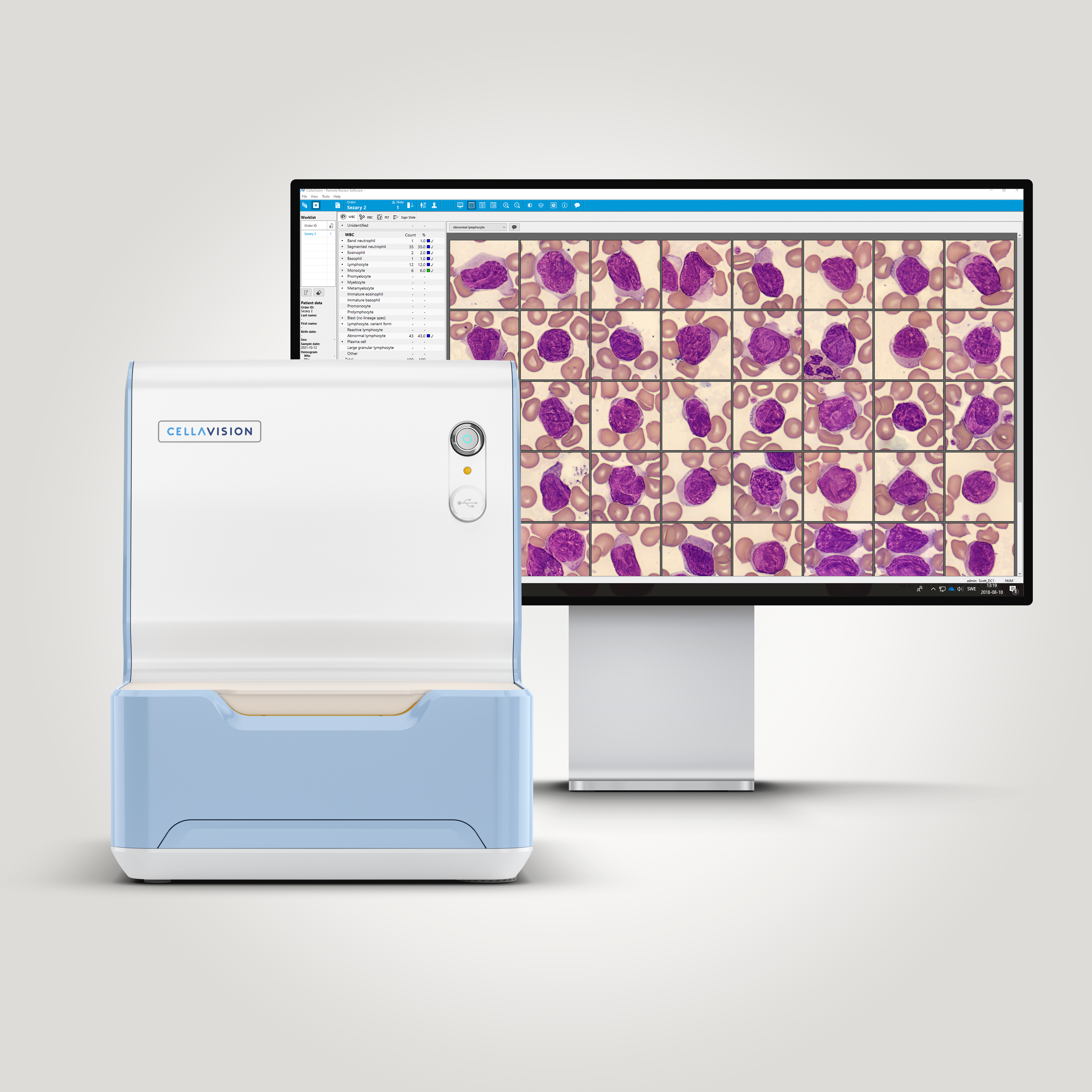
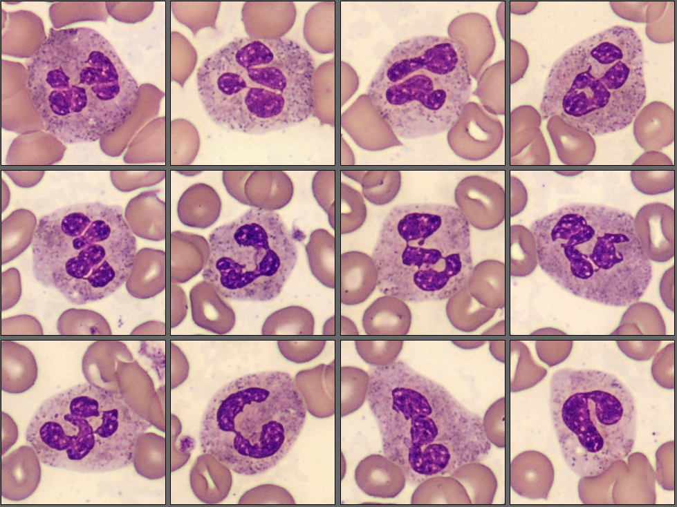
In the CellaVision® Remote Review, the lab technician examined the differential. What he saw was a normal population of neutrophils, lymphocytes and monocytes, but he also saw a large population among the lymphocytes which he re-classified to Abnormal lymphocytes.
Blood smear analysis on CellaVision® DC-1 Analyzer:
| WBC Differential | % | x109/L |
|---|---|---|
| Neutrophils | 36,9 | 3,4 |
| Eosinophils | 1,9 | 0,2 |
| Lymphocytes | 6,8 | 0,6 |
| Monocytes | 5,8 | 0,5 |
| Abnormal lymphocytes | 48,5 | 4,5 |
He then marked the sample for pathologist review and contacted the on-call hematopathologist. She opened the sample in the CellaVision® Remote Review Software and could see the normal populations of neutrophils, eosinophils, basophils, lymphocytes and monocytes.
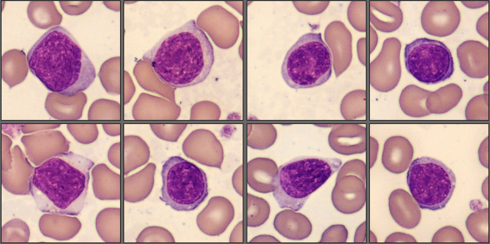
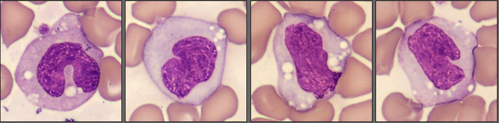
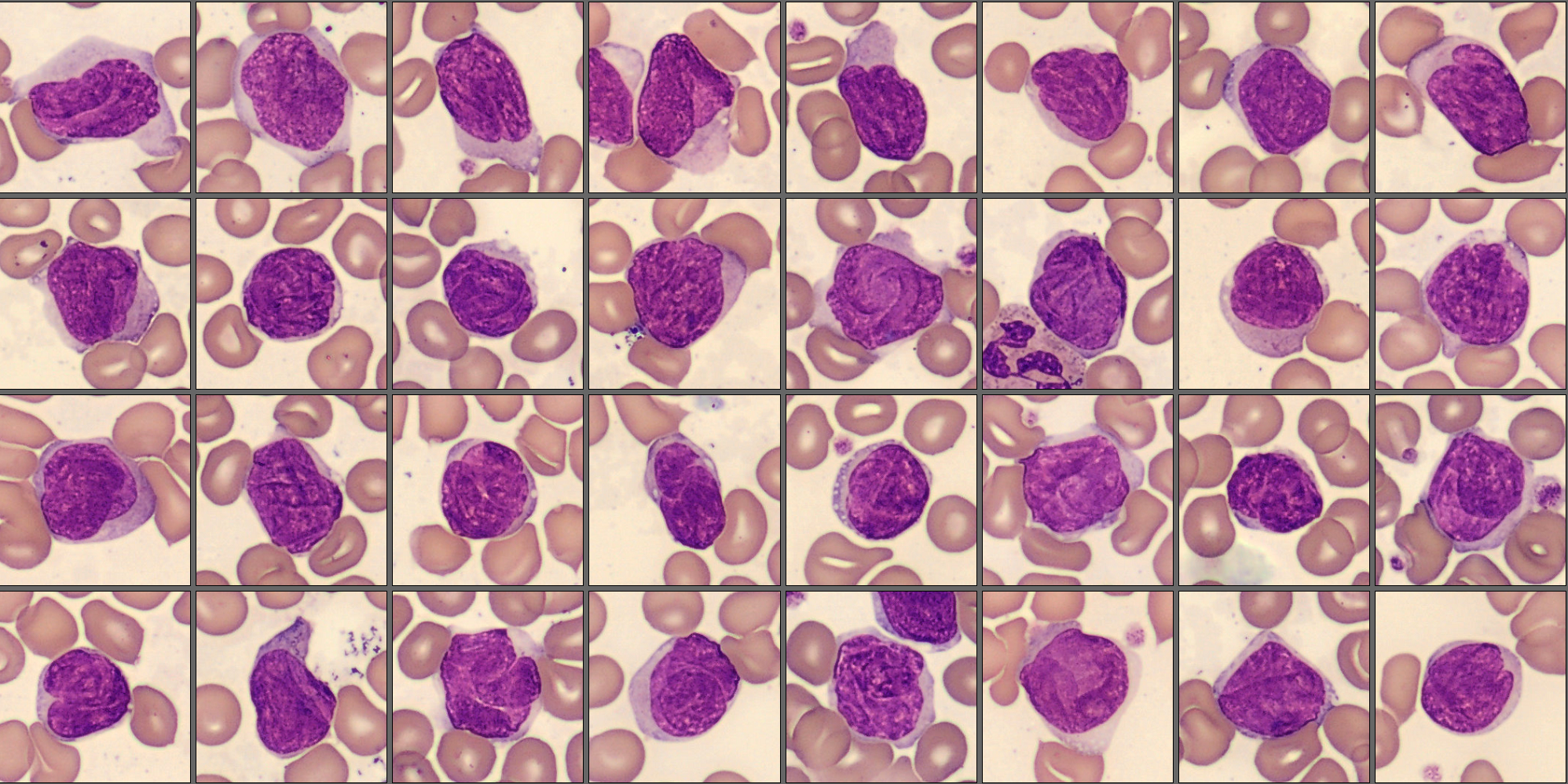
What she then saw in the Abnormal lymphocyte cell class was large lymphocyte-like cells with a large nucleus with a cerebriform shape.
To follow up, flow cytometry and cytogenetic tests were ordered.
Flow cytometry: CD2-, CD3+, CD4+, CD7-, CD8-, CD26-, CD27+, CD28+, CD45RO+
They also found deletions on chromosomes 10 and 17.
Diagnosis:
T Cell Lymphoma, Sézary Syndrome
Discussion:
Sézary Syndrome is a rare T-cell lymphoma which usually presents in patients >60+ years and is male predominant. Characteristically, it infiltrates the skin and can cause erythroderma (redness over most of the body) and alopecia (hair-loss). Many patients show lymphadenopathy and in more advanced cases, involvement in visceral organs.[1]
In the peripheral blood, it is typical to find large lymphoma cells with an abnormal nucleus with deep grooves and convolutions. These lymphoma cells can be small or large, but there can also be a mix of them. In the smaller cells, the cytoplasm is often scant while in the larger cells it is more visible. In the larger cells it is also easier to appreciate the cerebriform shape of the nucleus. The cytoplasm is often weakly basophilic and can contain vacuoles. [1][2]
Sézary syndrome is an aggressive disease and has a poor prognosis with a median survival time of 32 months. [1]
References:
[1] Elenitoba-Johnson, KSJ et. al. Sézary syndrome. In: WHO Classification of Tumors series, 5th ed.; vol 11, IARC publications, 2024. p. 674-676.
[2] Bain, Barbara J., (2015) Blood Cells, A Practical Guide (5th edition), John Wiley & Sons Ltd, p. 473-474; ISBN: 978-1-118-81733-9