Difference between Gram-positive and Gram-negative bacteria
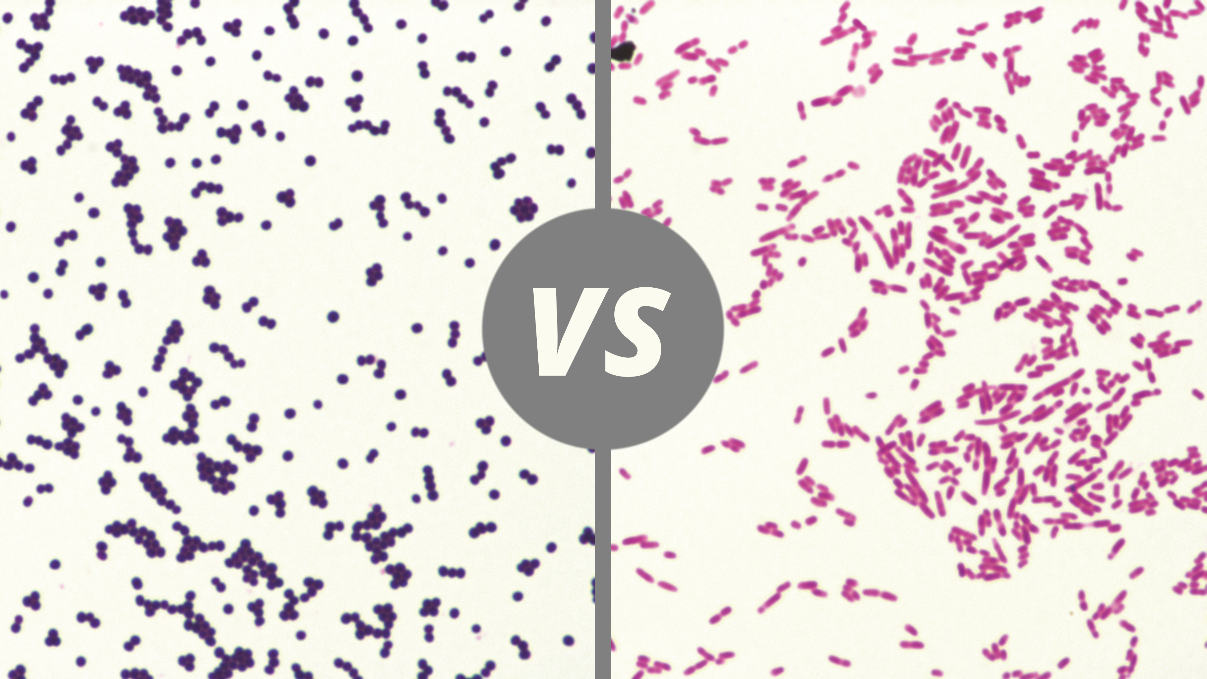
The classification of Gram-positive or negative bacteria relies on the appearance of the bacteria after a Gram staining.
What is Gram staining?
Named after Hans Christian Joachim Gram, Gram staining allows a differential staining of bacteria according to their cell wall composition.
Did you know? The initial Gram staining used gentian violet dissolved in Paul Ehrlich's solution and had no counterstain.
Over time, Gram staining was revisited to produce the two Gram staining methods that we know today, Gram Nicolle and Gram Hücker staining. The two staining’s differ mainly in regard to the stain and counterstains used.
The steps of Gram staining
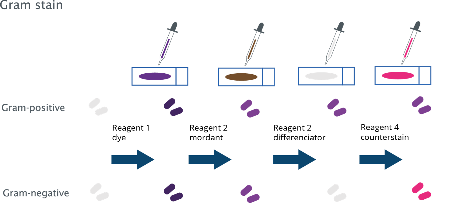
Gram staining is performed as follows:
- Stain a solution composed of gentian violet for a Gram Nicolle stain or a solution of Cristal violet for a Gram Hücker stain.
- Apply mordant, an iodine solution.
- Fade with alcohol/acetone.
- Counterstain with a fuchsin solution for Gram Nicolle stain or with a safranin solution for Gram Hücker stain.
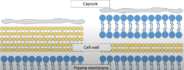
What causes the difference in bacteria staining?
| Gram positive | Gram negative |
|---|---|
| Inner most plasma membrane | Inner most plasma membrane |
| Thick peptidoglycan cell wall | Thin peptidoglycan cell wall |
| Outer capsule | Another plasma membrane |
| More easily treatable with antibiotics | Outer capsule |
| Stains purple/violet after Gram Stain | Harder to treat with antibiotics |
| Stains red/pink after Gram Stain |
The result of the Gram stain varies depending on the structure of the bacteria considered.
The general structure of the bacteria is:
- A plasma membrane made up of phospholipids, which regulate the entry and exit of the cell.
- A wall of peptidoglycans, covering the membrane and maintaining the cell’s structure.
Gram-positive and gram-negative bacteria differ in:
- The thickness of the peptidoglycan layer, which is thicker for Gram-positive than Gram-negative bacteria.
The thicker peptidoglycan layer in Gram-positive bacteria makes them resistant to alcohol/acetone decolorization, maintaining the violet staining known from Gram-positive bacteria brought by the first dye. Gram-negative bacteria are decolorized and then re-stained with the counterstain, producing a pink-orange staining depending on the Gram staining used. The counterstain added as Gram staining progresses makes it easier to visualize Gram-negative bacteria that have undergone alcohol/acetone decolorization