Patient case #1 - Sezary Syndrome
Description
A 67-year-old man presented with an extensive red, itchy rash on most of his body, unexplained weight loss, and lymphadenopathy. Testing showed an elevated AST, ALT, and LDH.
Hematology:
|
CBC result |
Result |
|
WBC |
9.3 x10⁹/L |
|
Hemoglobin |
12.6 g/dL |
|
Platelet Count |
165 x109/L |
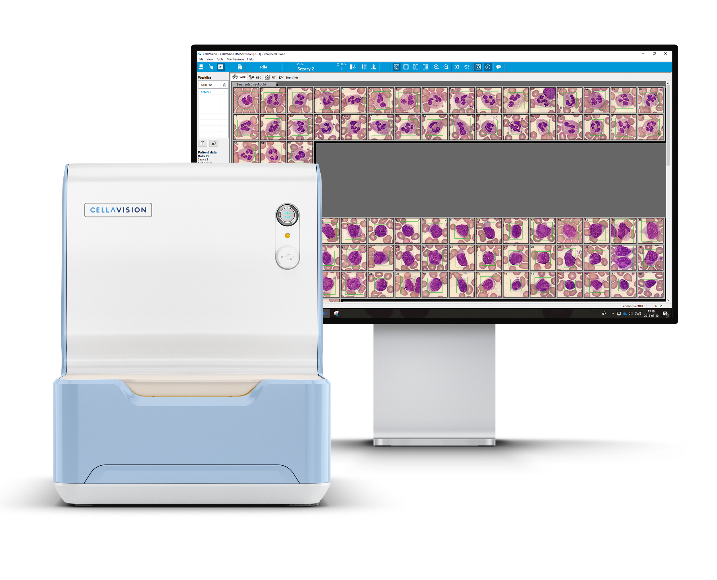
A “blasts?/abnormal lymph?” flag led to blood film analysis on Cellavision DC-1.
| WBC | % | x109/L |
|---|---|---|
| Neutrophils | 36.9 | 3.4 |
| Lymphocytes | 6.8 | 0.6 |
| Monocytes | 5.8 | 0.5 |
| Eosinophils | 1.9 | 0.2 |
| Others* | 48.5 | 4.5 |
| Smudge cells | 105 / 100 WBC |
* “Others” were described as large abnormal mononuclear cells with convoluted nuclei.
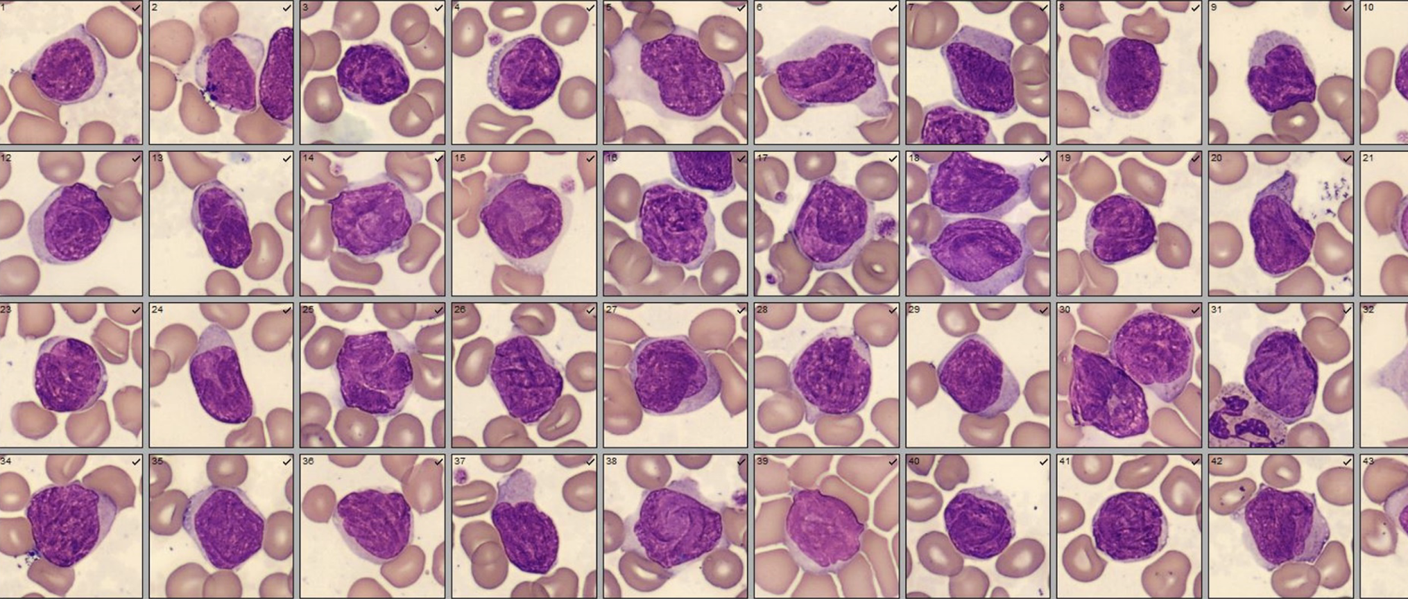
Side-by-side comparison of cell classes. (Cellavision DC-1, 100x, Wright-Giemsa stain).
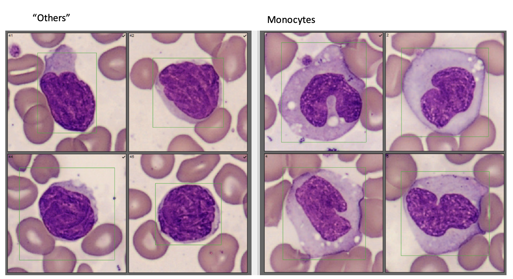
Side-by-side comparison of cells is helpful for differentiating between cell types. Images were taken with the Cellavision DC-1, 100x, Wright’s Giemsa stain. A peripheral blood sample was sent for flow cytometry testing. The “Other” cells were: CD2-, CD3+, CD4+, CD7-, CD8-, CD26-, CD27+, CD28+, CD45RO+.
Diagnosis: Sezary Syndrome
Sezary syndrome (SS) is a T cell lymphoma characterized by cutaneous and systemic involvement of Sezary cells in blood and lymph nodes. Typical Sezary cells are large with high nuclear : cytoplasmic ratio, convoluted, cerebriform nuclei, condensed chromatin and inconspicuous nucleoli. Some patients may exhibit smaller Sezary cells (Lutzner cells) which can be more difficult to recognize.